
Free data
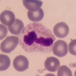
The requirement to access the firsthand data for the science specialist community is crystal clear to all. Meanwhile, collecting valid and labeled data by professionals, especially in the medical domain, is not an easy task. For instance, how many electrocardiograms or EEGs with the interpretation of prominent physicians are accessible for the science experts? Regarding the fact, Raabin health database attempts to facilitate providing and collecting the valid data in the medical domain on its behalf. Therefore, Raabin has the capacity to supply more than 40,000 microscopic images including 5 ranks of peripheral blood cells (neutrophil, eosinophil, basophil, lymphocyte, and monocyte) to the researchers for free in order to help them in the advancement of their research or commercial goals.
Raabin-WBC Data
Here you can choose and download the considered data by clicking on "related title" or "download here". Also, you can read more about the importance and necessity of WBC by clicking the title.
- Double-labeled Cropped Cells
- Nucleus_cytoplasm_Ground truths
- First Microscope Images
- Second Microscope Images
Double-labeled cropped cells are also provided containing only five main classes including mature neutrophils, lymphocytes (small and large), eosinophils, monocytes, and basophils. This sub-dataset is called Double-labeled Raabin-WB
Nucleus_cytoplasm_Ground truths
In recent years, many researchers have investigated segmenting the cytoplasm and nucleus of the white blood cells. Hence, we tried to prepare the ground truths of the cytoplasm and the nucleus for a proper number of cropped white blood cells. For this purpose, 1145 cropped images including 242 lymphocytes, 242 monocytes, 242 neutrophils, 201 eosinophils, and 218 basophils were randomly selected, and their ground truths were extracted by an expert.
dl.raabindata.com/WBC/nucleus_cytoplasm_GT/GroundTruths_bmpformat.rar
dl.raabindata.com/WBC/nucleus_cytoplasm_GT/GrTh.rar
dl.raabindata.com/WBC/nucleus_cytoplasm_GT/NucCytSplitter.py
Microscopic images were taken by the Olympus CX18 microscope and the Samsung Galaxy S5 camera
Note that the images of each blood film are placed in separate folders. Corresponding to each microscopic image, a dictionary (.json format) file containing the following information about that image was provided:
- Information about the blood elements in the image including their coordinates and labels.
- Information about the blood smears including staining method and the type of the disease.
- Information about the microscope includes the type of microscope and its magnification size.
- The type of camera used.
Microscopic images were taken by the Zeiss microscope and the LG G3 camera
Note that the images of each blood film are placed in separate folders. Corresponding to each microscopic image, a dictionary (.json format) file containing the following information about that image was provided:
- Information about the blood elements in the image including their coordinates and labels.
- Information about the blood smears including staining method and the type of the disease.
- Information about the microscope includes the type of microscope and its magnification size.
- The type of camera used.
Note that the images of each blood film are placed in separate folders. Corresponding to each microscopic image, a dictionary (.json format) file containing the following information about that image was provided:
- Information about the blood elements in the image including their coordinates and labels.
- Information about the blood smears including staining method and the type of the disease.
- Information about the microscope includes the type of microscope and its magnification size.
- The type of the camera used.
Here you can read the articles which has utilized Raabin-WBC Data:
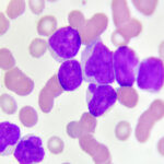
Raabin-Leukemia Data
Here you can choose and download the considered data by clicking on "related title" or "download here". Also, you can read more about leukemia by clicking the title.
- Acute Lymphoblastic Leukemia
- Acute Myeloblastic Leukemia
- Chronic Lymphocytic Leukemia
- Chronic Myelogenous Leukemia
All samples were taken from patients who had referred to our collaborator medical laboratory (Takht-e Tavous Laboratory in Tehran, Iran). It should be notices Zeiss microscope and LG J3 smartphone camera had been used for imaging.
All samples were taken from patients who had referred to our collaborator medical laboratory (Takht-e Tavous Laboratory in Tehran, Iran). It should be notices Zeiss microscope and LG J3 smartphone camera had been used for imaging.
All samples were taken from patients who had referred to our collaborator medical laboratory (Takht-e Tavous Laboratory in Tehran, Iran). It should be notices Zeiss microscope and LG J3 smartphone camera had been used for imaging.
All samples were taken from patients who had referred to our collaborator medical laboratory (Takht-e Tavous Laboratory in Tehran, Iran). It should be notices Zeiss microscope and LG J3 smartphone camera had been used for imaging.